
MRI has revolutionized sarcoma diagnosis and monitoring by providing detailed imaging of soft tissues. This non-invasive tool enables early detection, precise tumor assessment, and effective treatment planning. Post-treatment, MRI aids in evaluating therapy success, detecting recurrence, and ensuring comprehensive care. Advances in MRI technology continue to enhance sarcoma management, improving patient outcomes at every stage.
Understanding The Basics Of MRI and Its Applications In Sarcoma Cancer
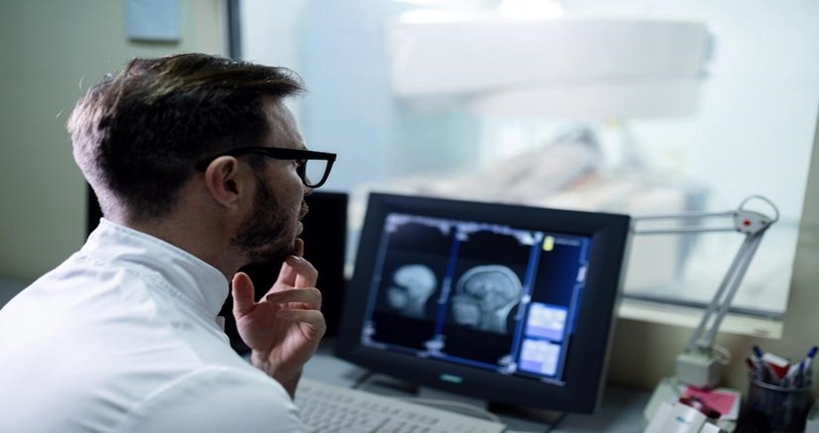
Magnetic Resonance Imaging (MRI) is a safe, non-invasive imaging method that uses magnetic fields and radio waves to produce detailed images of the body, avoiding ionizing radiation. Its ability to visualize soft tissues makes it invaluable for diagnosing sarcomas, which often originate in connective tissues. MRI provides high-resolution images, distinguishing healthy from abnormal tissue, and uses contrast agents to enhance tumor visibility. Beyond diagnosis, MRI is crucial for monitoring treatment, utilizing techniques like diffusion-weighted imaging to assess therapy response and guide further care. This versatility makes MRI indispensable in managing sarcoma patients.
How MRI Is Used For Diagnosing Sarcoma Cancer
Diagnosing sarcoma is challenging due to its rarity and nonspecific symptoms. MRI is an essential imaging tool in evaluating suspected cases, offering detailed images to distinguish benign from malignant tumors based on size, shape, and tissue characteristics. Radiologists identify potential malignancies by analyzing irregular margins, heterogeneous signals, and tissue infiltration. Multi-planar imaging provides a comprehensive view of tumor anatomy, aiding disease assessment and treatment planning. Additionally, MRI guides biopsy procedures by pinpointing the optimal site for tissue sampling, enhancing diagnostic accuracy, and reducing complications.
The Importance Of MRI In Staging And Treatment Planning For Sarcoma Cancer
Accurate sarcoma cancer staging is crucial for effective treatment planning, and MRI plays a pivotal role by providing detailed insights into tumor size, location, and involvement of nearby structures. This information helps determine whether the cancer is localized or has metastasized, guiding classification systems.
In treatment planning, MRI is instrumental in defining surgical margins, ensuring complete tumor removal while preserving healthy tissue. It also evaluates the tumor’s response to neoadjuvant therapies, such as chemotherapy or radiation, by monitoring changes in size and characteristics. These capabilities make MRI essential for tailoring and optimizing sarcoma cancer treatment strategies, enhancing the effectiveness of the overall management plan.
MRI’s Role In Monitoring Treatment Response And Assessing Tumor Progression
To detect recurrence and evaluate therapy effectiveness, post-treatment monitoring is essential for sarcoma patients. MRI provides high-resolution images that reveal subtle changes in tumor size or characteristics. Advanced techniques like diffusion-weighted imaging help distinguish viable tumor tissue from necrotic or fibrotic tissue, offering insights into tumor behavior and treatment response. Routine MRI scans can identify asymptomatic recurrences early, enabling timely interventions that improve prognosis and outcomes. This proactive approach enhances survival rates and quality of life for sarcoma patients.
Advancements In MRI Technology For Sarcoma Cancer Imaging
Advancements in MRI technology have significantly improved its role in sarcoma imaging. High-field MRI systems (3 Tesla or higher) provide superior image clarity, revealing intricate tumor details and surrounding tissue. Functional MRI and spectroscopy further enhance diagnostic capabilities by assessing blood flow, metabolic activity, and biochemical profiles of tumors, aiding in treatment planning and response evaluation.
Integrating artificial intelligence (AI) into MRI analysis transforms sarcoma care, enabling faster, more accurate image interpretation. AI enhances the detection of subtle anomalies, streamlines diagnostics, and supports timely, effective treatment decisions.
Potential Limitations And Challenges Of MRI In Sarcoma Cancer Diagnosis And Monitoring
While MRI offers significant sarcoma diagnosis and monitoring advantages, it faces particular challenges. Limited availability in rural or under-resourced areas can delay diagnosis and treatment, highlighting the need for equitable access. Accurate interpretation of MRI findings is another concern, as distinguishing between benign and malignant sarcomas requires specialized expertise. Ongoing training and collaboration between radiologists and oncologists are crucial to reduce misdiagnoses. Additionally, MRI may be less effective for evaluating bone involvement or distant metastases, necessitating a multimodal imaging approach, including CT or PET scans, for comprehensive assessment and treatment planning.
Integrating MRI With Other Imaging Modalities For Comprehensive Sarcoma Cancer Care
Integrating MRI with other imaging modalities optimizes sarcoma management by leveraging their complementary strengths. MRI excels in soft tissue visualization, and combining it with CT scans enhances the assessment of bone lesions and metastatic disease due to CT’s superior imaging of bony structures. PET scans evaluate metabolic activity and identify active disease sites through increased glucose uptake. When paired with MRI, PET provides a comprehensive view of tumor viability and treatment response.
Emerging hybrid systems like PET/MRI combine anatomical and metabolic data in a single scan, offering a detailed, holistic perspective of the tumor’s behavior. This multimodal approach improves diagnostic accuracy, refines treatment planning, and supports effective monitoring of disease progression. At Tellica Imaging, these advanced imaging techniques deliver comprehensive insights that enhance sarcoma management and treatment outcomes.
Promising Research And Future Directions For MRI In Sarcoma Cancer Management
Ongoing research enhances MRI’s role in sarcoma care by developing targeted contrast agents to better detect small lesions. Artificial intelligence (AI) and machine learning advances improve pattern recognition and outcome prediction, reducing image interpretation variability and increasing diagnostic accuracy. Researchers are also exploring MRI’s potential to assess tumor microenvironments and identify biomarkers related to treatment response. Techniques to visualize changes in tumor vasculature, cellularity, and metabolism could inform personalized treatment strategies, improving outcomes for sarcoma patients.
Conclusion: The Growing Significance Of MRI in Sarcoma Cancer Care
MRI is pivotal in sarcoma care, from diagnosis to post-treatment monitoring. It offers high-resolution, noninvasive imaging crucial for clinical decision-making and patient outcomes. As advancements in technology and research progress, MRI’s capabilities in diagnosing and managing sarcoma are set to expand further, enhancing precision and treatment options.
Integrating MRI with other imaging modalities and artificial intelligence holds promise for personalized and comprehensive care. Collaborative efforts among radiologists, oncologists, and researchers are vital to maximizing MRI’s potential, ensuring patients receive optimal outcomes and improved quality of life in their cancer journey.
Comments are closed.